√無料でダウンロード! eye of tiger sign mri 623423-Eye of tiger mri
These disorders are classified based on genetic differences The most common NBIA disorder is autosomal recessive pantothenate kinaseassociated neurodegeneration (PKAN) PKAN is commonly associated with the eyeofthetiger sign;Older individuals may exhibit dementia and ambulation is eventually impaired The MRI usually shows an area of hyperintensity in the medial globus pallidus that has been called the 'eye of the tiger' sign but this is not pathognomonic Axonal degeneration with accumulation of spheroidal inclusions can be seen histologicallyThe most wellknown hallmark of the syndrome is the eyeofthetiger sign on the brain magnetic resonance imaging (MRI) scan Previous studies have highlighted a onetoone correlation between the MRI findings of the eyeofthetiger sign and the presence of a pantothenate kinase 2 ( PANK2 ) mutation, postulating that the MRI appearance is a good diagnostic tool for identifying PANK2 mutationpositive cases

The Eye Of The Tiger Sign Radiology
Eye of tiger mri
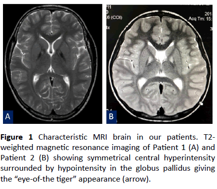
Eye of tiger mri-Abstract Pantothenate kinaseassociated neurodegeneration (PKAN) is a rare disorder associated with brain iron accumulation The brain MRI abnormality consists of T2 hypointensity in the globus pallidus with a small hyperintensity in its medial part, called the "eyeofthetiger" sign We report on 2 patients affected by PKAN, in whom MRI examination did not demonstrate the "eyeofthetiger" sign in the early stages;The socalled eyeofthetiger sign is a hallmark for PKAN This sign consists of hypointense areas of iron deposits that surround a region of hyperintense signal due to gliosis in both globus pallidus on T2weighted images 14


Q Tbn And9gcqdhd4xpivj6ydrphfz5kf Nktr4vr4fwibufy58 Usqp Cau
A range of other brain MR imaging changes have been reported in patients with this diagnosis, including the pattern designated as the eyeofthetiger sign, which combines high signal intensity in the center of the globus pallidus interna with low signal intensity in the surrounding region 2 We have shown that there is an absolute correlation between the presence of a mutation in PANK2 and the eyeofthetiger sign 3;The eye of the tiger sign (EOT), seen on T2 sequences of magnetic resonance imaging (MRI), is considered pathognomonic for neurodegeneration with brain iron accumulation type 1 (NBIA1, previously known as Hallervorden–Spatz syndrome) 1–3This sign is described as low signal intensity in both globi pallidi (due to iron accumulation) that surrounds a central region of high signal intensityEndometriomas, also known as chocolate cysts or endometriotic cysts, are a localized form of endometriosis and are usually within the ovary They are readily diagnosed on ultrasound, with most demonstrating classical radiographic features Epid
Funduscopy confirmed the finding of RP, and an MRI showed marked bilateral highsignal intensities surrounding the globus pallidus — the "eye of the tiger" sign that is characteristic of pantothenate kinaseassociated neurodegeneration (PKAN) (Fig 1 left;A particular change, called the eyeofthetiger sign, which indicates an accumulation of iron, is typically seen on magnetic resonance imaging (MRI) scans of the brain in diseaseinfosearchorg Due to the accumulation of iron in the basal ganglia, two black spots can be seen, which is referred to as the " eye of the tiger " signFunduscopy confirmed the finding of RP, and an MRI showed marked bilateral highsignal intensities surrounding the globus pallidus — the "eye of the tiger" sign that is characteristic of pantothenate kinaseassociated neurodegeneration (PKAN) (Fig 1 left;
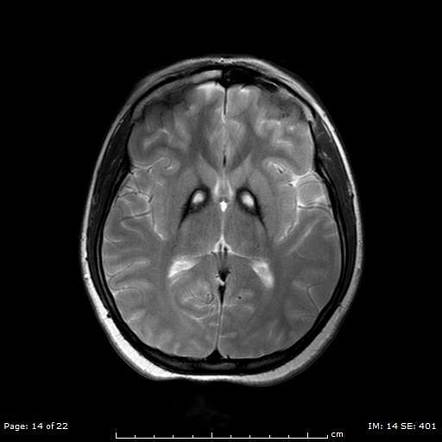
Eye of the tiger sign is seen in Hallervorden Spatz syndrome, also known as pantothenate kinaseassociated neurodegeneration ( PKAN ) This sign describes characteristic low signal surrounding a central region of high signal in the anteromedial globus pallidus on T2 Pathologically, the low signal corresponds to abnormal iron deposition, and the high signal corresponds to gliosis, vacuolisation and increased water contentIron deposition in conjunction with destruction of the globus pallidus gives rise to the characteristic eyeofthetiger sign in MRI It has been postulated that pantothenate kinase 2 mutations underlying all cases of classic HallervordenSpatz syndrome are always associated with the eyeofthetiger signAn eye‐of‐the‐tiger sign is a specific magnetic resonance imaging (MRI) pattern, a key diagnostic feature of pantothenate kinase associated neurodegeneration (PKAN) It is low‐signal intensity rings surrounding the central high‐signal intensity regions in the medial aspect of bilateral globus pallidus on T2‐weighted MRI The surrounding hypointensity of the globus pallidus is due to excess iron accumulation


Figure 1 From Mr Image Mimicking The Eye Of The Tiger Sign In Wilson S Disease Semantic Scholar


Cureus Eye Of The Tiger Sign In Neurodegeneration With Brain Iron Accumulation
The right image shows an agematched normal MRI) 1Eye Tiger Sign A sign seen on T2weighted MRI, in which there is a very low signal intensity in the globus pallidus due to an excess accumulation of iron, surrounding a central region of high signal intensity attributed to gliosis, increased water content and neuronal loss with disintegration, vacuolization and cavitation of the neuropilThe most wellknown hallmark of the syndrome is the eyeofthetiger sign on the brain magnetic resonance imaging (MRI) scan Previous studies have highlighted a onetoone correlation between the MRI findings of the eyeofthetiger sign and the presence of a pantothenate kinase 2 (PANK2) mutation, postulating that the MRI appearance is a good


Eye Of The Tiger Sign In Hallervorden Spatz Syndrome Eurorad


Eye Of The Tiger A Key Diagnostic Sign Of Pantothenate Kinase Associated Neurodegeneration Eurorad
Iron deposition in conjunction with destruction of the globus pallidus gives rise to the characteristic eyeofthetiger sign in MRI It has been postulated that pantothenate kinase 2 mutations underlying all cases of classic HallervordenSpatz syndrome are always associated with the eyeofthetiger signEye of the tiger sign is seen in Hallervorden Spatz syndrome, also known as pantothenate kinaseassociated neurodegeneration ( PKAN ) This sign describes characteristic low signal surrounding a central region of high signal in the anteromedial globus pallidus on T2 Pathologically, the low signal corresponds to abnormal iron deposition, and the high signal corresponds to gliosis, vacuolisation and increased water contentWe describe the eyeofthetiger sign on magnetic resonance imaging (MRI) of the brain in a 40yearold man presenting with extra pyramidal symptoms like chorea, flexion neck dystonia, tongue tremors, dysarthria and postural instability as the sequelae of organophosphorus poisoning six months previously This typical radiological sign has been described in extrapyramidal parkinsonian disorders including corticalbasal ganglionic degeneration, early onset levodoparesponsive parkinsonism and


Dr Balaji Anvekar Frcr Hallervorden Spatz Syndrome
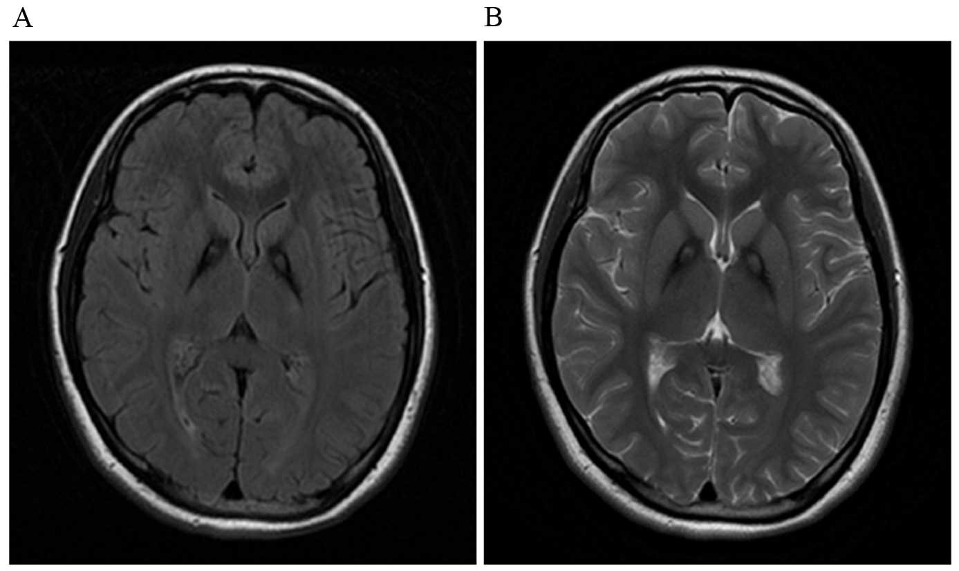


Novel Homozygous Pank2 Mutation Identified In A Consanguineous Chinese Pedigree With Pantothenate Kinase Associated Neurodegeneration
The most wellknown hallmark of the syndrome is the eyeofthetiger sign on the brain magnetic resonance imaging (MRI) scan Previous studies have highlighted a onetoone correlation between the MRI findings of the eyeofthetiger sign and the presence of a pantothenate kinase 2 (PANK2) mutation, postulating that the MRI appearance is a good diagnostic tool for identifying PANK2 mutationpositive casesAnd abnormal postures, movements, and tremors If other family members are also affected, this may help determine the diagnosisMRI brain in our patient had shown hyperintense streaking of the globus pallidus in the region of the medial medullary lamina and surrounding hypointensity suggesting the "eye of tiger sign" mimic This MRI finding has been described in a series of patients with MPAN


Leopard Skin Sign White Matter Radiology Reference Article Radiopaedia Org


View Image
Magnetic resonance imaging (MRI) of brain demonstrates a characteristic 'eyeofthetiger' sign We describe a case of NBIA in a child with classical clinical and MRI of brain featuresThe right image shows an agematched normal MRI) 1EYE OF THE TIGER sign is the MRI changes seen in Globus Pallidus in Pantothenate kinase associated neurodegenration The Globus Pallidus in T2W MRI shows medial high signal and lateral low signalPantothenate kinase associated neurodegenrationMost common form of neurodegeneration with iron accumulation in the brainMutation of pantothenate kinase 2 gene is the etiologyBest imaging manifestation


T2w Axial A And Coronal B Mri Images Show The Classic Download Scientific Diagram


The Eye Of The Tiger Sign Cmaj
T2weighted magnetic resonance images (MRI) showed bilaterally marked hypointensity with a central region of hyperintensity in the globus pallidus, or the socalled "eyeofthetiger" signWe describe the eyeofthetiger sign on magnetic resonance imaging (MRI) of the brain in a 40yearold man presenting with extra pyramidal symptoms like chorea, flexion neck dystonia, tongue tremors, dysarthria and postural instability as the sequelae of organophosphorus poisoning six months previouslyThe eyeofthetiger signb allows the specific MR imaging diagnosis of HallervordenSpatz syndrome or related extrapyramidal parkinson disorders in the presence of supporting clinical signs


Mri Findings Nbia


Looking Deep Into The Eye Of The Tiger In Pantothenate Kinase Associated Neurodegeneration American Journal Of Neuroradiology
MRI brain in our patient had shown hyperintense streaking of the globus pallidus in the region of the medial medullary lamina and surrounding hypointensity suggesting the "eye of tiger sign" mimic This MRI finding has been described in a series of patients with MPANT2weighted magnetic resonance images (MRI) showed bilaterally marked hypointensity with a central region of hyperintensity in the globus pallidus, or the socalled "eyeofthetiger" signThe brain MRI findings, the patient's history and physical examination findings, along with the similar family history allowed a suggestion of Hallervorden Spatz syndrome However, the eye of the tiger sign is suggestive but not pathognomonic of this entity A trial of carbidopa did not show noticeable improvement of her spasticity


Figure 1 From Pantothenate Kinase 2 Mutation With Classic Pantothenate Kinase Associated Neurodegeneration Without Eye Of The Tiger Sign On Mri In A Pair Of Siblings Semantic Scholar


Eye Of The Tiger Sign Idi Medicos Adda Facebook
We describe the eyeofthetiger sign on magnetic resonance imaging (MRI) of the brain in a 40yearold man presenting with extra pyramidal symptoms like chorea, flexion neck dystonia, tongue tremors, dysarthria and postural instability as the sequelae of organophosphorus poisoning six months previously This typical radiological sign has been described in extrapyramidal parkinsonian disorders including corticalbasal ganglionic degeneration, early onset levodoparesponsive parkinsonism andSurprisingly, magnetic resonance imaging revealed a typical Eye of the Tiger sign within the basal ganglia ( Fig 1 ) A mutation in the PANK2 gene was excluded via genetic testing 1 The MR scan of our patient with multiple system atrophy shows the Eye of the Tiger Sign in the striatum (left and middle) and reduced availability of Dopamin D2 receptors on IBZM‐SPECT (right – coregistration with MRI)The eyeofthetigersign is known as a radiological sign, that refers to abnormal low signal intensity in the globus pallidus, with a central longitudinal zone of high signal, as seen on T2weighted MRI images In the transverse plane through the basal ganglia this appears as a tiger like image with prominent eyes


T2 Weighted Brain Mri Of The 39 Year Old Patient Showed Bilateral Download Scientific Diagram


Classic Pkan Nbia
We describe the eyeofthetiger sign on magnetic resonance imaging (MRI) of the brain in a 40yearold man presenting with extra pyramidal symptoms like chorea, flexion neck dystonia, tongue tremors, dysarthria and postural instability as the sequelae of organophosphorus poisoning six months previouslyFor instance, in one study, 100% of the examined PKAN cases had an eyeofthetiger sign on brain MRI A genetic panel for NBIA, specifically PKAN, was ordered for this patient including pantothenate kinase 2 (PanK2) the genetic marker for pantothenate kinaseEYE OF THE TIGER sign is the MRI changes seen in Globus Pallidus in Pantothenate kinase associated neurodegenrationT The Globus Pallidus in T2W MRI shows medial high signal and lateral low signal Pantothenate kinase associated neurodegenration Most common form of neurodegeneration with iron accumulation in the brain


Typical Brain Mri In A Patient With Pkan Showing Eye Of The Tiger Sign Download Scientific Diagram


Eye Of The Tiger Sign In T2 Image Figure 4 Eye Of Tiger Sign In Download Scientific Diagram
Iron deposition in conjunction with destruction of the globus pallidus gives rise to the characteristic eyeofthetiger sign in MRI It has been postulated that pantothenate kinase 2 mutations underlying all cases of classic HallervordenSpatz syndrome are always associated with the eyeofthetiger signRecently, a few PSP cases have reported the "eye of the tiger" sign on MRI examinations The "eye of the tiger" sign, in globus pallidus, is a sign that bilaterally symmetrically located low signal intensity and central longitudinal hyperintensity are observedThe typical abnormalities were detected only in the following examinations


T2 And Fse Mri Distinguishes Four Subtypes Of Neurodegeneration With Brain Iron Accumulation Neurology


Clinical Reasoning A 16 Year Old Boy With Freezing Of Gait Neurology
The eye of the tiger sign refers to abnormal low T2 signal on MRI (due to abnormal accumulation of iron) in the globus pallidus with a longitudinal stripe of high signal (due to gliosis and spongiosis) The eye of the tiger sign is most classically associated with pantothenate kinaseassociated neurodegeneration 13 although it is not pathognomonic 5Cortex is usually spared but caudate atrophy may be seen in more advanced cases The eye of the tiger sign refers to a central T2 relatively hyperintense spot (line) within the hypointense globi pallidi due to gliosis and vacuolisation 3Presence of the Eyeofthetiger Sign on Magnetic Resonance Imaging in a Subject with Atypical HallervordenSpatz Syndrome Lacking Pantothenate Kinase 2 Mutation


View Image
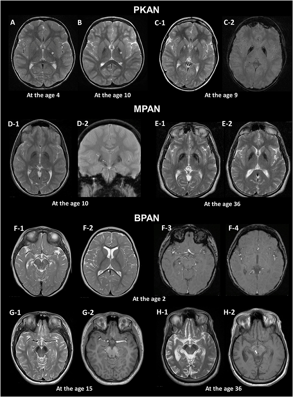


Frontiers Brain Mri Pattern Recognition In Neurodegeneration With Brain Iron Accumulation Neurology
We describe the eyeofthetiger sign on magnetic resonance imaging (MRI) of the brain in a 40yearold man presenting with extra pyramidal symptoms like chorea, flexion neck dystonia, tongue tremors, dysarthria and postural instability as the sequelae of organophosphorus poisoning six months previouslyThe eye of the tiger sign refers to a central T2 relatively hyperintense spot (line) within the hypointense globi pallidi due to gliosis and vacuolisation 3 MR spectroscopy shows decreased NAA peak due to neuronal loss and may show increased myoinositol 8;MRI image shows iron deposits in the basal ganglia, the socalled eyeofthetiger sign (T2w GRASE sequence) A neurological examination would show evidence of muscle rigidity;


Medical Eponyms With Explanations 12


Mri Of The Brain T2 Weighted Sequence Eye Of The Tiger Sign A T2 Download Scientific Diagram
The face of the giant panda sign in neuroimaging refers to the appearance of the midbrain, when the red nucleus and substantia nigra are surrounded by high T2 signal in the tegmentum It is classically seen in Wilson disease, although whenever the white matter is diffusely abnormal in the region a similar appearance will be perceived such as in Japanese encephalitisDetails Dear Sirs, The eye of the tiger sign (EOT), seen on T2 sequences of magnetic resonance imaging (MRI), is considered pathognomonic for neurodegeneration with brain iron accumulation type 1 (NBIA1, previously known as Hallervorden–Spatz syndrome) 1 – 3 This sign is described as low signal intensity in both globi pallidi (due to iron accumulation) that surrounds a central region of high signal intensity (caused by gliosis, edema, and neuronal loss with subsequent secondaryThat is, all patients with the PANK2 mutation have this MR imaging


Stereotactic Pallidotomy In A Child With Hallervorden Spatz Disease In Journal Of Neurosurgery Volume 90 Issue 3 1999


Figure 3 From Pantothenate Kinase 2 Mutation With Classic Pantothenate Kinase Associated Neurodegeneration Without Eye Of The Tiger Sign On Mri In A Pair Of Siblings Semantic Scholar
The right image shows an agematched normal MRI) 1This sign consists of hypointense areas of iron deposits that surround a region of hyperintense signal due to gliosis in both globus pallidus on T2weighted images 14 In rare cases, the eyeofthetiger sign is not found in patients with PKAN, although presymptomatic diagnosis of the disease by means of MRI findings has been described 1The characteristic imaging finding includes the 'eyeofthetiger' sign which refers to a specific pattern of signal alteration in the globus pallidus that is seen on T2weighted MR images This refers to symmetrical low signal intensity circumscribing a central region of high signal intensity in the globus pallidus on either side


Magnetic Resonance Imaging In Pantothenate Kinase 2 Associated Neurodegeneration


Pantothenate Kinase 2 Mutation With Classic Pantothenate Kinase Associated Neurodegeneration Without Eye Of The Tiger Sign On Mri In A Pair Of Siblings Semantic Scholar
Funduscopy confirmed the finding of RP, and an MRI showed marked bilateral highsignal intensities surrounding the globus pallidus — the "eye of the tiger" sign that is characteristic of pantothenate kinaseassociated neurodegeneration (PKAN) (Fig 1 left;Hayflick et al reported a onetoone correlation between the magnetic resonance imaging (MRI) findings of the eyeofthetiger sign and the presence of a PANK2 mutation and postulated that the MRI appearance is a good diagnostic tool for identifying PANK2 mutationpositive cases 2 Herein, we report an atypical HSS patient without a PANK2That is, all patients with the PANK2 mutation have this MR imaging
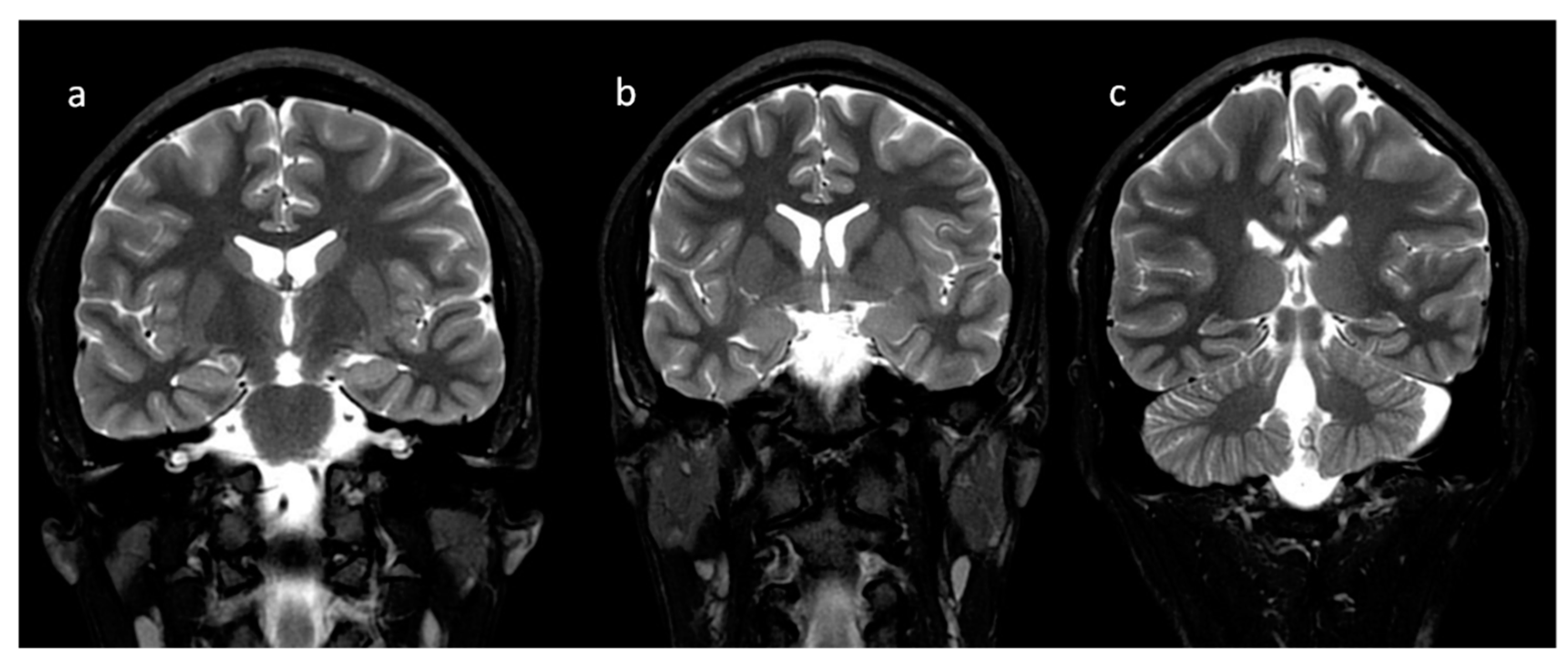


Brain Sciences Free Full Text Neuroimaging Of Basal Ganglia In Neurometabolic Diseases In Children Html


A Diagnostic Approach For Neurodegeneration With Brain Iron Accumulation Clinical Features Genetics And Brain Imaging
A range of other brain MR imaging changes have been reported in patients with this diagnosis, including the pattern designated as the eyeofthetiger sign, which combines high signal intensity in the center of the globus pallidus interna with low signal intensity in the surrounding region 2 We have shown that there is an absolute correlation between the presence of a mutation in PANK2 and the eyeofthetiger sign 3;Recently, a few PSP cases have reported the "eye of the tiger" sign on MRI examinations The "eye of the tiger" sign, in globus pallidus, is a sign that bilaterally symmetrically located low signal intensity and central longitudinal hyperintensity are observedThe socalled eyeofthetiger sign is a hallmark for PKAN This sign consists of hypointense areas of iron deposits that surround a region of hyperintense signal due to gliosis in both globus pallidus on T2weighted images 14


Magnetic Resonance Imaging Susceptibility Weighted Imaging And Quantitative Susceptibility Mapping Findings Of Pantothenate Kinase Associated Neurodegeneration Sciencedirect


Http Pdf Posterng Netkey At Download Index Php Congress Ecr15 Module Get Pdf By Id Poster Id
Pantothenate kinase associated neurodegeneration (PKAN) is the most prevalent type of neurodegeneration with brain iron accumulation (NBIA) disorders characterized by extrapyramidal signs, and 'eyeofthetiger' on T2 brain magnetic resonance imaging (MRI) characterized by hypointensity in globus pallidus and a hyperintensity in its core


Kjr Korean Journal Of Radiology


Neuroacanthocytosis A Rare Movement Disorder With Magnetic Resonance Imaging Topic Of Research Paper In Clinical Medicine Download Scholarly Article Pdf And Read For Free On Cyberleninka Open Science Hub


Looking Deep Into The Eye Of The Tiger In Pantothenate Kinase Associated Neurodegeneration American Journal Of Neuroradiology
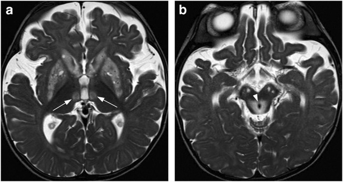


Bilateral Lesions Of The Basal Ganglia And Thalami Central Grey Matter Pictorial Review Springerlink


Journals Sagepub Com Doi Pdf 10 1177
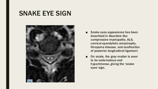


Eye Signs In Radiology


Eye Of The Tiger Sign In T2 Weighted Brain Mri Download Scientific Diagram


A Neurological Mri Menagerie Practical Neurology
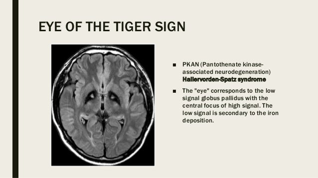


Eye Signs In Radiology
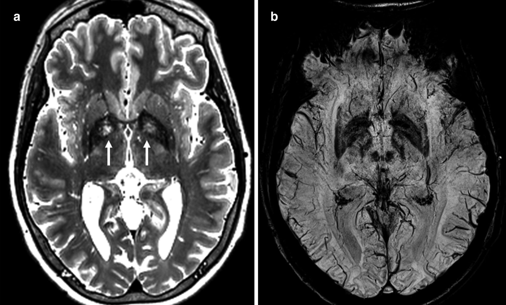


Brain Springerlink
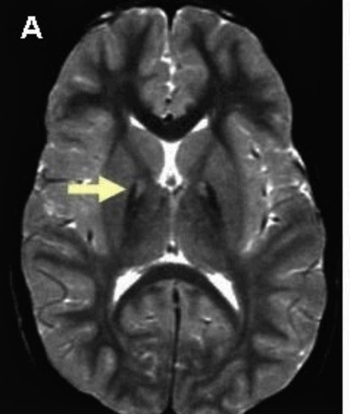


Pantothenate Kinase Associated Neurodegeneration Medlineplus Genetics


Q Tbn And9gcqdhd4xpivj6ydrphfz5kf Nktr4vr4fwibufy58 Usqp Cau


Neurodegeneration With Brain Iron Accumulation Nbia Eurorad


Vinod


As Iron Goes So Goes Disease Haematologica


Eye Of The Tiger A Key Diagnostic Sign Of Pantothenate Kinase Associated Neurodegeneration Eurorad


The Role Of Genetic Mutations In Gene Pank2 On Hallervorden Spatz Syndrome


Www Japi Org R2f4d474 Hallervorden Spatz Disease
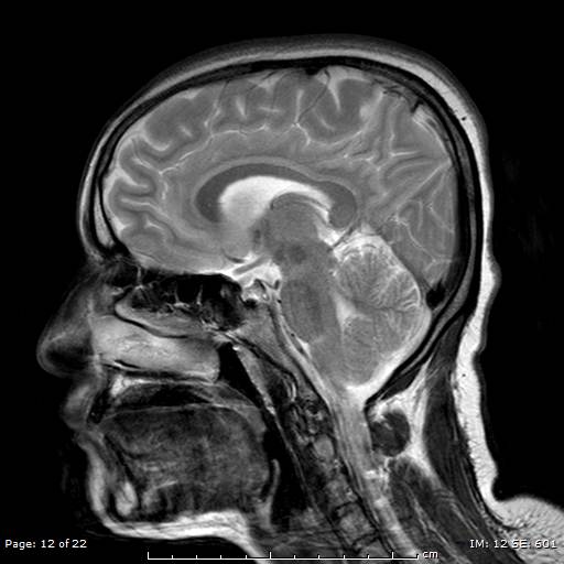


Hallervorden Spatz Syndrome Eye Of The Tiger Sign Image Radiopaedia Org


Q Tbn And9gcrgxxc9hrcpzv7wc3akd7olryxj0eew01mnfrqtlmxabkdjlogs Usqp Cau


Kjr Korean Journal Of Radiology


Ajns African Journal Of Neurological Sciences Pantothenate Kinase Associated Neurodegeneration Case Series


Thejcn Com Synapse Data Pdfdata 0145jcn Jcn 5 192 Pdf


A Diagnostic Approach For Neurodegeneration With Brain Iron Accumulation Clinical Features Genetics And Brain Imaging


Epos Trade


Http Www Ijars Net Articles Pdf 2377 Ce Vsu F Ang Pf1 Vsu Ang Pfa Ang Pb Vsu Ang Pn Ang Pdf


Frank Gaillard Who S A Pretty Kitty Then Excellent Example Of Eye Of The Tiger Sign T Co Vzctirtnbn Mri Radiology Brain


Eye Of The Tiger Sign And Classic Pantothenate Kinase Associated Neurodegeneration Springerlink
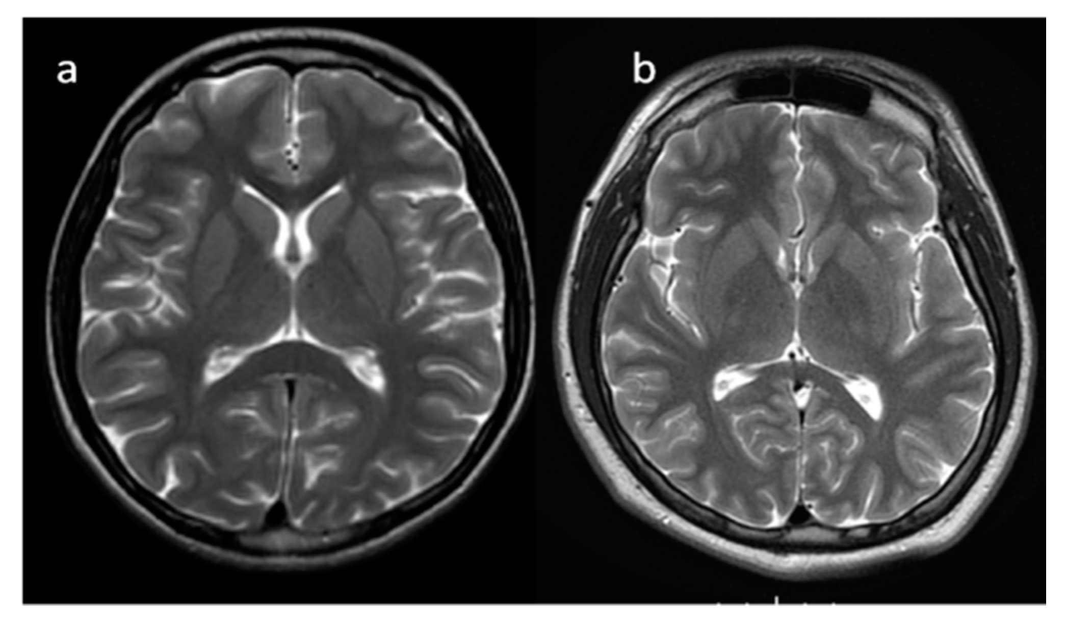


Brain Sciences Free Full Text Neuroimaging Of Basal Ganglia In Neurometabolic Diseases In Children Html


Dementia E Neuropsychologia


Brain Mri In Neurodegeneration With Brain Iron Accumulation With And Without Pank2 Mutations American Journal Of Neuroradiology


The Eye Of The Tiger Sign Radiology


Viewing Playlist aa Julypin Radiopaedia Org Pediatric Radiology Radiology Brain Images


Late Onset Atypical Pantothenate Kinase Associated Neurodegeneration
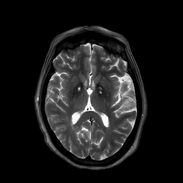


Hallervorden Spatz Syndrome Eye Of The Tiger Sign Radiology Case Radiopaedia Org


Eye Of The Tiger Sign In A 48 Year Healthy Adult Sciencedirect


Brain Mri In Neurodegeneration With Brain Iron Accumulation With And Without Pank2 Mutations American Journal Of Neuroradiology


Neuroimaging Features Of Neurodegeneration With Brain Iron Accumulation American Journal Of Neuroradiology


Pantothenate Kinase Associated Neurodegeneration Pkan


A Diagnostic Approach For Neurodegeneration With Brain Iron Accumulation Clinical Features Genetics And Brain Imaging
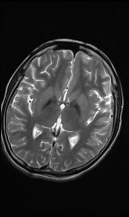


Eye Of The Tiger Sign Globus Pallidus Radiology Reference Article Radiopaedia Org


Progressive Brain Iron Accumulation In Neuroferritinopathy Measured By The Thalamic T2 Relaxation Rate American Journal Of Neuroradiology


Cureus Eye Of The Tiger Sign In Neurodegeneration With Brain Iron Accumulation


Epos Trade


Eye Of The Tiger A Key Diagnostic Sign Of Pantothenate Kinase Associated Neurodegeneration Eurorad


Two Siblings With Atypical Pantothenate Kinase Associated Neurodegeneration


Neuroimaging Features Of Neurodegeneration With Brain Iron Accumulation American Journal Of Neuroradiology


Clinical Applications Chapter 17 Magnetic Resonance Imaging In Movement Disorders


Molecular Analysis Of Pank2 Gene In Two Thai Classic Pantothenate Kinase Associated Neurodegeneration Pkan Patients Insight Medical Publishing


Http Pdf Posterng Netkey At Download Index Php Module Get Pdf By Id Poster Id


Eye Of The Tiger Sign Globus Pallidus Radiology Reference Article Radiopaedia Org


Axial T2 Weighted Mri Image Showing Eye Of The Tiger Sign With Download Scientific Diagram


Mri Findings Nbia


Q Tbn And9gcssgkbolkwl3itvcl7c8iv 7umbmbtmz1hbi8uyqx1rf1bexx Usqp Cau


Movement Disorders Role Of Imaging In Diagnosis Mascalchi 12 Journal Of Magnetic Resonance Imaging Wiley Online Library


Q Tbn And9gctdk9pjq3wnquryj8xmg1yew9z Yyzaia5kektavdcdpccvhj36 Usqp Cau


Http Pdf Posterng Netkey At Download Index Php Module Get Pdf By Id Poster Id


The Eye Of The Tiger Brain Iron And The Beta Propeller Beyond The Ion Channel


Pank2 Gene An Overview Sciencedirect Topics


Axial T2 Weighted Mri Image Showing Eye Of The Tiger Sign With Download Scientific Diagram


Toxic And Metabolic Brain Disease Radiology Key


Eye Of The Tiger Sign In A 23year Patient With Mitochondrial Membrane Protein Associated Neurodegeneration Journal Of The Neurological Sciences


Hallervorden Spatz Disease Instagram Posts Photos And Videos Picuki Com


Hallervorden Spatz Syndrome Eye Of The Tiger Sign Radiology Case Radiopaedia Org


T2w Axial A And Coronal B Mri Images Show The Classic Download Scientific Diagram


Tiger Eye Sign Tyger Tyger Burning Bright Radioman


Mri Findings Nbia


Movement Disorders Role Of Imaging In Diagnosis Mascalchi 12 Journal Of Magnetic Resonance Imaging Wiley Online Library
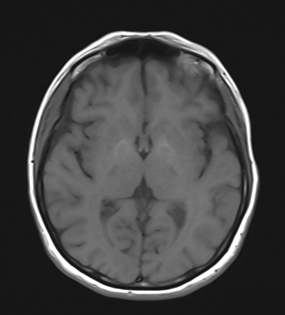


Eye Of The Tiger Sign Radiology Case Radiopaedia Org


Movement Disorders Role Of Imaging In Diagnosis Mascalchi 12 Journal Of Magnetic Resonance Imaging Wiley Online Library


Patterns Of Brain Iron Accumulation Ppt Download


コメント
コメントを投稿